Fluorescence Detection of Membrane Potential
Creative Bioarray can complete a full range of cell electrophysiological testing and analysis. We have established a complete experimental protocol, relying on ion indicators and potential probes for ion concentration analysis and membrane potential detection of cultured cells. This service can help customers with corresponding research needs quickly complete analysis and testing and harvest high-quality data.
Membrane Potential Imaging Method
The cytoplasmic membrane separates the internal environment of the cell from the external environment, allowing nutrients, ions, biomolecules, and genetic material to accumulate, which is a necessary condition for the existence of life on earth. A particularly important aspect of membrane biology is the ability to separate ions of different concentrations and types. The resulting membrane potential is the basic biophysical property maintained by every cell on the earth. The increase and decrease of membrane potential play a central role in many physiological processes. The coordination and rapid changes of membrane potential realize many advanced functions such as nerve impulse transmission and muscle contraction.
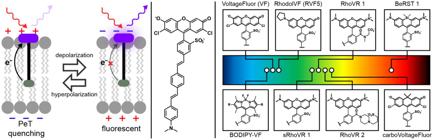 Fig 1. A glowing and growing palette of Voltage-sensitive Fluorophores (VoltageFluors). (Liu P, et al. 2019)
Voltage imaging has gradually become an important technique to reveal the impact of rapid changes in membrane voltage on cell physiology. Small molecule voltage imaging has a long history, and compounds with voltage-sensitive optical properties provide a shortcut for the fluorescence detection of membrane potential. Traditional voltage-sensitive dye molecules are divided into two categories: "fast" dyes and "slow" dyes. "Fast" dyes are usually electrochromic dyes that can be used to monitor rapid changes in neuronal voltage. At the same time, a variety of ion indicators such as calcium ion indicators can also realize the visualization of membrane potential through the movement of ions across the membrane.
Service Based on Ion Indicators
This service uses many ion indicators to track calcium and other ion concentrations. We provide imaging applications based on calcium indicator, sodium indicator, potassium indicator, magnesium indicator, zinc indicator and other ionic indicators to meet the research needs of different customers. According to the cell sample commissioned or submitted by the customer and the research purpose, select the appropriate ion indicator, and track the concentration of calcium and other ions through a strong fluorescent signal and a series of wavelength options. This service can provide customers with some commercial indicators, as well as customized services based on reactive fluorescent dyes. You only need to feedback the samples and requirements to us, and we will provide you with one-stop analysis and testing services.
 Fig 2. Resolvable ion type.
Service Based on Membrane Potential Indicators
We provide membrane potential changes analysis services for nerve cells, cardiomyocytes, and other cell types based on membrane potential probes. This service can be targeted at clients with research needs, such as nerve impulse propagation, myocardial contraction, and cell signaling. We select industry-leading potential probes to analyze and test cell samples commissioned or submitted by customers. The core of this service is slow or fast response detection. The fast response probe can be used for preliminary analysis of physiological processes with instantaneous (millisecond) potential changes, such as nerve impulses. In contrast, slow probes can help to explore the mitochondrial function and cell viability.
Application
- For the ion concentration of tissue and cell samples, see Monitoring and Electrical Activity Imaging;
- Quantitative analysis of membrane potential changes in response to pharmacological stimuli;
- Track the changes of some ion concentrations in organelles and vesicle structures;
Creative Bioarray uses industry-leading ion indicators and potential probes to track the concentration of target ions in cells and analyze membrane potential. From commercial ion indicators to customized reactive fluorescent dye-labeled probes, our services can meet the diverse needs of customers and help you achieve satisfactory results with minimal cost. Our scientific team will provide you with the solution that best suits your needs. If you are interested in our services or have any specific needs, please contact us. We look forward to working with you in the near future.
References:
- Liu P, Miller E W. Electrophysiology, unplugged: imaging membrane potential with fluorescent indicators[J]. Accounts of chemical research, 2019, 53(1): 11-19.
- Miller E W. Small molecule fluorescent voltage indicators for studying membrane potential[J]. Current opinion in chemical biology, 2016, 33: 74-80.
For research use only. Not for any other purpose.