Cell Phagocytosis and Transport Function Assay Services
Creative Bioarray provides comprehensive cell transmembrane transport analysis services. Based on live cell imaging technology and a variety of fluorescent labeling dyes and indicators, we realize the analysis of cell phagocytosis, endocytosis, and internalization. Advanced flow sorting system and confocal imaging system helps researchers to achieve high-resolution visual analysis needs. Our platform has established experimental protocols for a variety of analysis methods and can provide suitable solutions according to the researcher's sample type and analysis needs.
Phagocytosis & Phagocytosis
Cellular material transport "crosses" the plasma membrane in different ways according to the nature, size, and function of the transferred material. In the transport of non-nutrient substances, more attention has been paid to phagocytosis (phagocytes, antigen presenting cells), endocytosis, and ligand-mediated transport of substances across membranes. Pathogen clearance and antigen presentation of phagocytes existing in the immune system cannot be separated from the help of phagocytosis. The phagocytosis of target particles involves coordinated steps of recognition, binding to phagocyte receptors, and internalization. Endocytosis is the process of endocytosis of non-protein or polysaccharides and other non-particulate materials. Imaging and quantification of the various stages of phagocytosis help to clarify the molecular mechanisms of this cellular process.
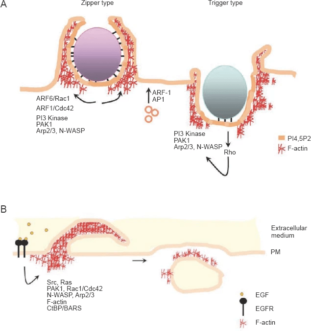 Fig 1. Macroscale endocytic processes. (Kumari S, et al. 2010)
Cell Phagocytosis and Transport Function Assay
Our cell phagocytosis and material transport analysis service aims to use a series of fluorescent dyes and indicators to visualize the process of cell transmembrane material transport. Our experimental platform has advanced confocal imaging and flow sorting systems, and has established a complete imaging analysis experimental protocol to help customers complete visual analysis.
- Phagocytosis. The principle is to use fluorescent dye-labeled indicators (processed yeast or bacteria) to detect and track phagocytosis, which can detect simple ingestion processes. This process can use confocal imaging equipment for image recording.
- Endocytosis. The principle is to use fluorescent protein and organic dyes to dye the vesicle wall and vesicle content. Or through the principle that the pH of the vesicle changes during endocytosis, the pH indicator is used to detect the entire process of the pathway.
- Ligand/antibody internalization. The principle is to use common receptors as targets for fluorescent ligand-mediated live cell imaging. Multiple indicators based on indicators can be used to track the movement of specific targets in multiple membrane compartments. In addition, this service can also use fluorescently labeled antibodies for target analysis through a flow cytometry system.
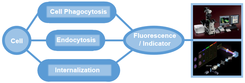 Fig 2. Visual detection services related to phagocytosis and internalization.
Creative Bioarray provides cell visualization analysis services related to cell phagocytosis and internalization. We use advanced confocal imaging and flow cytometry equipment, using a variety of fluorescent dyes and indicators to complete quantitative analysis. Through the use of live cell imaging technology, high-resolution analysis of cell phagocytosis, endocytosis, and ligand-mediated internalization is carried out. If you are interested in our services or have related analysis needs, please feel free to contact us. We look forward to working with you in the near future.
References:
- Kumari S, Swetha M G, Mayor S. Endocytosis unplugged: multiple ways to enter the cell[J]. Cell research, 2010, 20(3): 256-275.
- Rashidfarrokhi A, Richina V, Tafesse F G. Visualizing the early stages of phagocytosis[J]. Journal of visualized experiments: JoVE, 2017 (120).
- Smirnov A, Solga M D, Lannigan J, et al. Using imaging flow cytometry to quantify neutrophil phagocytosis[M]//Neutrophil. Humana, New York, NY, 2020: 127-140.
For research use only. Not for any other purpose.