Exosomal Staining and Tracking Services
Creative Bioarray provides technical services for exosome staining and visual tracking analysis. This service helps researchers understand the process of visualizing the life cycle of exosomes and obtain accurate spatiotemporal assessments of exosomes. The application of advanced imaging equipment and live cell imaging technology to visualization of exosomes has promoted the development of exosomes research. With our exosomes service experience and expertise, we can provide customers with satisfactory exosomes visualization analysis solutions.
Exosomes Tracking and Visualization
Extracellular vesicles (EVs) are nano-scale vesicles released from cells with powerful autocrine and paracrine biological activities. Small extracellular vesicles called exosomes affect a variety of autocrine and paracrine cell phenotypes. Although EVs were considered to be cell fragments with little biological relevance in the early days, more and more evidences show that EVs constitute the basic mode of cell-to-cell communication, which can mediate specific protein, nucleic acid and lipid cargoes in different environments. Delivery of recipient cells. In fact, EVs are involved in the pathogenesis of many diseases, including infections, neurodegenerative diseases, cardiovascular diseases, and cancers. Understanding the function of exosomes requires multiple tools, including real-time imaging. Exosomal staining combined with fluorescent labels can visualize the exosomal life cycle, including multivesicular body (MVB) transport, MVB fusion, exosomal uptake, and endosome acidification. The accurate spatiotemporal assessment of exosomes and their contents can be visualized and analyzed by specific and powerful live cell imaging technology and imaging equipment.
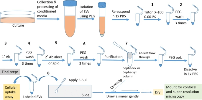 Fig 1. Strategy for labeling of Extracellular Vesicles (EV) derived from glioma cells. (Mondal A, et al. 2019)
Exosomes Staining and Visualized Exosome TrackingServices
Understanding the transport or movement of exosomes in biological systems will provide researchers with clear visual evidence to interpret the functions of exosomes and the specific uptake selectivity of exosomes. The visualization of exosomes in biological samples requires analysis of exosomes after staining. Reagents and tools used to isolate, characterize and analyze their RNA and protein content, as well as in vitro and in vivo tracking, can help researchers further understand exosomes. Many labeling techniques can be used to characterize exosomes, such as the use of metabolic incorporation of modified nucleic acids or methionine analogs. This method may allow more in-depth interrogation of the RNA and protein content of a given vesicle. Our exosomal staining service can use fluorescent dyes for selective labeling of exosomal RNA and membrane components. This service is aimed at in vitro visual analysis (separated exosomes samples), and can also help with in vivo verification of exosomes tracing.
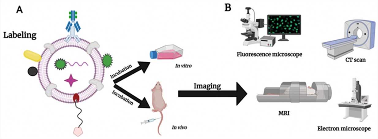 Fig 2. Illustration of labeling exosomes with different probes and NPs, and different instruments that can be used to image exosomes in vitro and in vivo. (Hamzah R N, et al. 2021)
Labeling strategy. We provide a variety of terminal inspection equipment, such as fluorescence microscopes (conventional confocal fluorescence microscopes, high-resolution fluorescence microscopes), electron microscopes (SEM and TEM), flow cytometers, etc. Depending on the detection equipment, our exosomes labeling methods mainly use commercial magnetic beads, fluorescent antibodies and artificial nanoparticles.
Target type. The structure of exosomes is a lipophilic spherical structure, and its surface and content contain nucleic acids, proteins and some lipids.
Creative Bioarray has a complete exosome analysis equipment platform. With many years of experience in providing exosomes research products and services, you can rely on our expertise to meet all your exosomes visualization analysis needs. If you are interested in our services or have any specific needs, please contact us. We look forward to working with you in the near future.
References:
- Mondal A, Ashiq K A, Phulpagar P, et al. Effective visualization and easy tracking of extracellular vesicles in glioma cells[J]. Biological procedures online, 2019, 21(1): 1-12.
- Hamzah R N, Alghazali K M, Biris A S, et al. Exosome Traceability and Cell Source Dependence on Composition and Cell-Cell Cross Talk[J]. International Journal of Molecular Sciences, 2021, 22(10): 5346.
For research use only. Not for any other purpose.