Creative Bioarray has extensive experience in providing our customers with high-quality 3D tumor spheroid assays to accelerate their research and drug development.
Introduction of 3D Tumor Spheroid Assays
Solid tumor is a complex three-dimensional (3D) structure composed of specific tissues and cells. Tumor cells are an important part of tumor specific cytoarchitecture. The complex interactions within solid tumors and with the environment affect the characteristics of tumor cells, such as gene expression and protein distribution. Many in vitro models lack a tumor-related physiological environment to truly evaluate the response of tumor cells to external stimuli. The conventional two-dimensional (2D) culture models have limited value in tumor research. Thus, 3D culture methods have been developed to establish tumor model systems with higher physiological relevance, so as to better mimic the heterogeneity and complexity of tumors.
In 3D culture models, the growth of tumor cells usually forms multicellular aggregates or spheroids. The cancer stem/progenitor cells can be enriched and proliferated to form tumor spheres by culturing tumor cells on ultra-low adherent surface in serum-free media supplemented with growth factors, such as epidermal growth factor (EGF) and basic fibroblast growth factor (bFGF). Compared to cell monolayers, 3D tumor spheroid models provide a more physiological tissue-like morphology and close cell-cell contacts to examine physiological properties of tumor cells.

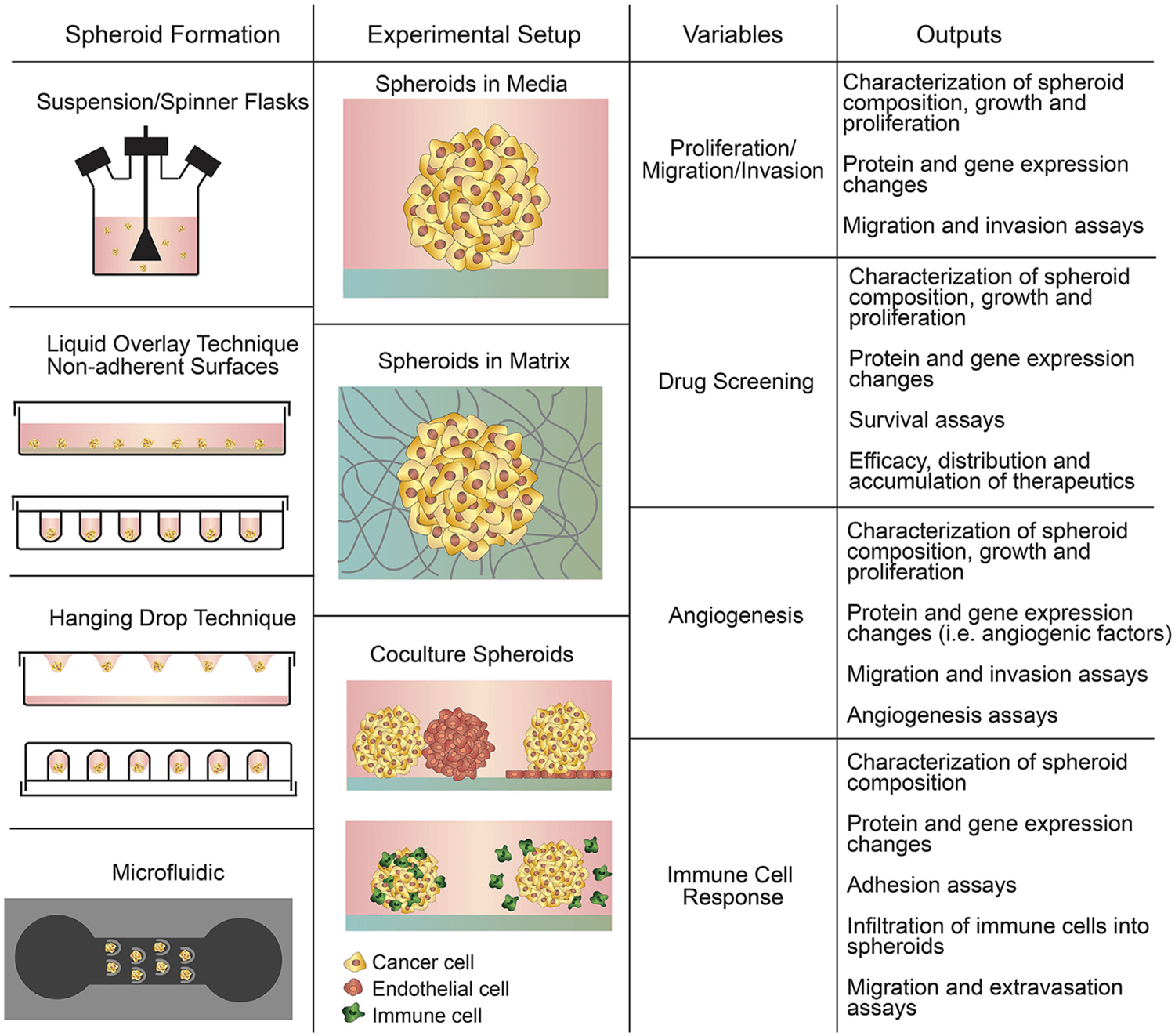
Figure 1. Summary of spheroid-based assays including spheroid formation techniques, experimental setups, variables to study, and experimental outputs. (Katt M E, et al., 2016)
3D tumor spheroid assays have become a powerful and important tool to assess the viability, proliferation, invasion, migration and angiogenesis of tumor cells. They also have emerged as advanced tools to accelerate drug discovery efforts. 3D culture models provide a reliable, standardized, flexible, and high-throughput compatible platform for basic research and anti-cancer drug screening.

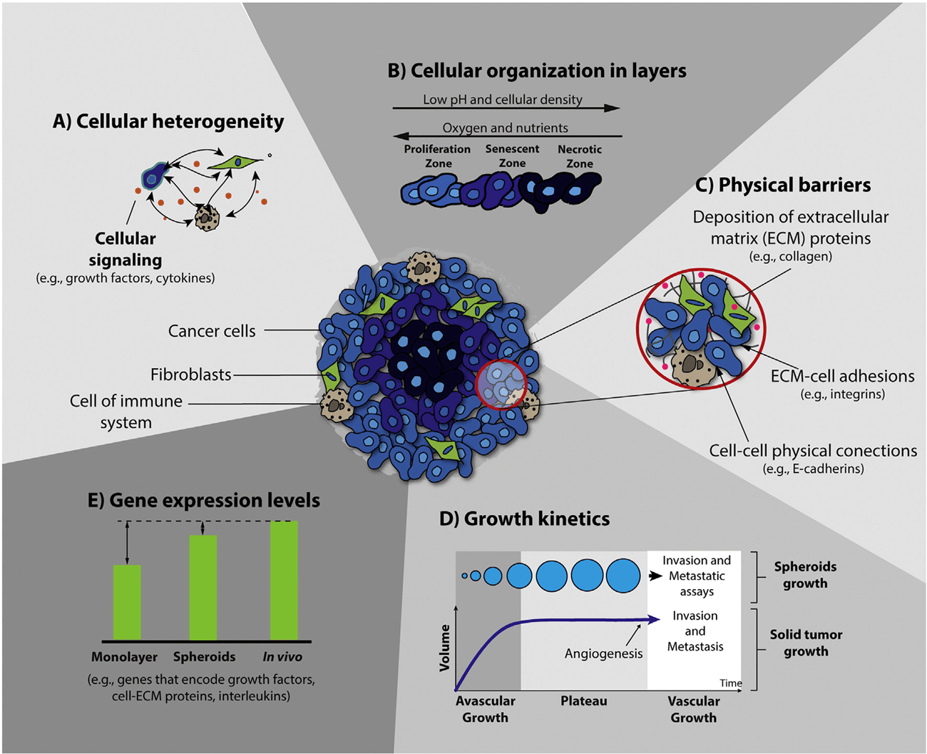
Figure 2. Schematic representation of the main characteristics of 3D spheroids that are crucial for their application in the screening of anticancer therapeutics. (Costa E C, et al., 2016)
Applications of 3D Tumor Spheroid Assays
- Used to study cell function in tumor microenvironment.
- Used to study the pathophysiologic pathways of tumors.
- Used to analyze the self-renewal ability of cancer stem cells (CSCs).
- Used for CSC enrichment.
- Used to characterize and estimate the percentage of cancer stem/progenitor cell population.
- Used to analyze the effect of tested compounds on mono-spheroids of tumor cells or co-culture spheroids of tumor and stroma cells.
- Used for anti-cancer small-molecule screening.
- Used to predict the potential efficacy and clinical trial outcomes of candidate drugs.
- Used as a promising innovative strategy for future cancer therapy.
Advantages of 3D Tumor Spheroid Assays
➢ Offer more physiologically relevant information of in vivo tumors.
- Physiological morphology.
- Microenvironment, including cell-cell interactions, cell-ECM interactions and immune interactions.
- Different nutritional and oxygen status.
- Tumorigenesis, signaling transduction, metastasis, and cancer progression.
- Tumor heterogeneity.
➢ Compatible with high-throughput, high-content, real-time and automation techniques.
➢ More translatable into anti-tumor activity in vivo.
3D Tumor Spheroid Assays Available at Creative Bioarray
For tumor research and drug screening, we can provide both scaffold-based and scaffold-free methods to determine the formation of mono-spheroids of tumor cells or co-spheroids of tumor and stroma cells.
Scaffold-Based Methods
The scaffolds can support the proliferation of tumor cells and promote cell interactions. The commonly used scaffolds include synthetic hydrogels, ECM-based natural hydrogels and engineered hydrogels. Spheroids can be seeded on top of or inside the matrix. Spinner flasks, bioreactors, micropatterned plates and microfluidic devices can also be used to create tumor spheroids.
Scaffold-Free Methods
Scaffold-free methods used to generate tumor spheroids generally include ultra-low cell attachment plates, hanging drop, magnetic levitation and 3D printing. These methods are suitable to culture tumor cells which can spontaneously self-aggregation into highly organized 3D structures.
Creative Bioarray has experienced scientific team working closely with our customers to provide high-quality customize services with fast turnaround time. If you need more detailed information, please feel free to contact us. We look forward to cooperating with you.
References:
- Katt M E, et al. In vitro tumor models: advantages, disadvantages, variables, and selecting the right platform. Frontiers in bioengineering and biotechnology, 2016, 4: 12.
- Costa E C, et al. 3D tumor spheroids: an overview on the tools and techniques used for their analysis. Biotechnology advances, 2016, 34(8), pp: 1427-1441.
For research use only. Not for any other purpose.


