Exosomes Identification Services
Creative Bioarray provides exosome identification services for biological samples and purified exosomes samples. This service provides a variety of method-based solutions to facilitate researchers with different testing purposes. Our platform has established a variety of mature protocols to support your exosome research, from isolation to characterization.
Exosomes Identification
The characterization of exosomes has specificity and identity, so multiple characterization indicators are needed to determine whether the extracted components are exosomes. The International Association of Extracellular Vesicles (ISEV) recommends evaluating two types of proteins (eg transmembrane or GPI-anchored proteins associated with plasma membrane and/or endosomes, cytoplasmic proteins recovered from extracellular vesicles (EV)) To determine whether the extracted components are exosomes, the purity of exosomes derived from biological fluids can be assessed by determining the presence or absence of certain non-EV structural protein components co-separated with EV. Since the selection of different purification methods leads to the persistence of different cellular contaminants (cell debris or other proteins, lipids, etc.) during the purification process, exosomes characterization technology is very important in the field of exosomes.
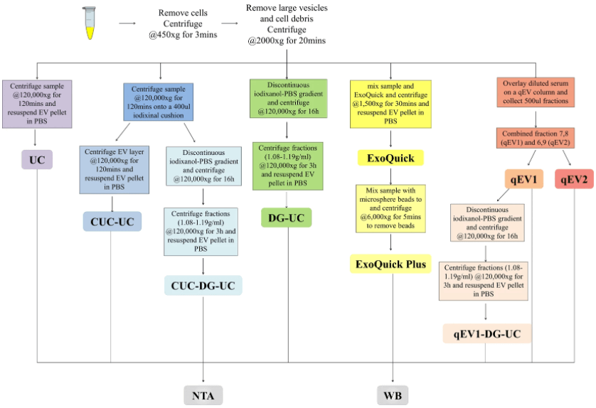 Fig 1. Schematic summary of EV isolation and downstream analyses. EVs were isolated from human serum using five different methods alone or in combination and characterized by Western blot (WB), nanoparticle tracking analysis (NTA). (Brennan K, et al. 2020)
Exosomes Characterization Service Based on Multiple Methodologies
Exosomes characterization methods are mainly divided into two categories: external characterization (mainly morphology and particle size detection) and inclusion body characterization (such as membrane proteins, lipid rafts). In general research, researchers can choose to consider the identification of isolated exosomes from three different levels, including transmission electron microscope (TEM) identification of exosomes morphology, nanoparticle tracking analysis (NTA) identification of exosomes size, and exosomal surface proteins Western Blot identification of markers.
Morphological Characterization of Exosomes
We provide visualization and characterization services of exosomes based on electron microscopy. Identify the components of exosomes in samples or after purification by providing high-resolution microscopic imaging equipment. Electron microscopy imaging can directly see the morphology of the particles, so it can be used to identify the presence and integrity of exosomes. Exosomal characterization services through electron microscopy can not only distinguish smaller-sized particles, but also observe cup-shaped vesicles whose morphology and size are compatible with extracellular vesicles. This direct visualization method is very useful for the quality identification of exosomes after isolation.
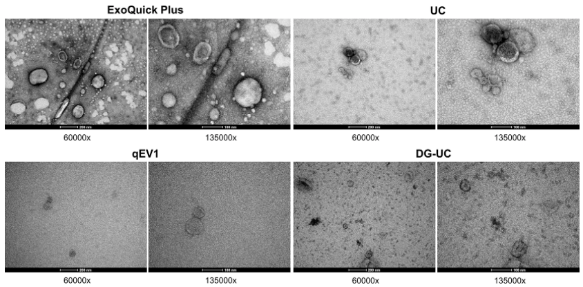 Fig 2. Transmission electron microscopic (TEM) visualization of the EVs isolated from pooled human serum. (Brennan K, et al. 2020)
Exosome Size Characterization
We provide services for analyzing the size and concentration of exosomes based on NTA technology. The advantage of this technology is its fast detection speed and real-time observation of exosomes. Our NAT analysis is mainly for samples after separation and purification. The platform has NTA-related equipment for characterizing 10nm-1000nm nanoparticles in solution. Each particle is individually but simultaneously analyzed by direct observation and measurement of diffusion events. In addition to measuring particle size and concentration, the fluorescence mode provides differentiation of intrinsic or fluorescently labeled nanoparticles.
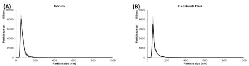 Fig 3. Nanosight analysis of particle size distribution of the EVs isolated from pooled human serum. (Brennan K, et al. 2020)
Identification of Protein Markers on the Surface of Exosomes
Our exosomes indicate that the service of protein marker identification can be carried out through various methods such as western blotting, ELISA, and flow cytometry. These methods can be selected according to different detection purposes. Western blotting can perform qualitative and quantitative analysis of labeled proteins, and it is easier to analyze exosomes from cell culture media. ELISA also has the characteristics of strong specificity and rapid detection and can perform qualitative and quantitative analysis of labeled proteins. Flow cytometry has high-throughput, multi-channel analysis capabilities, fast analysis speed, and low sample concentration.
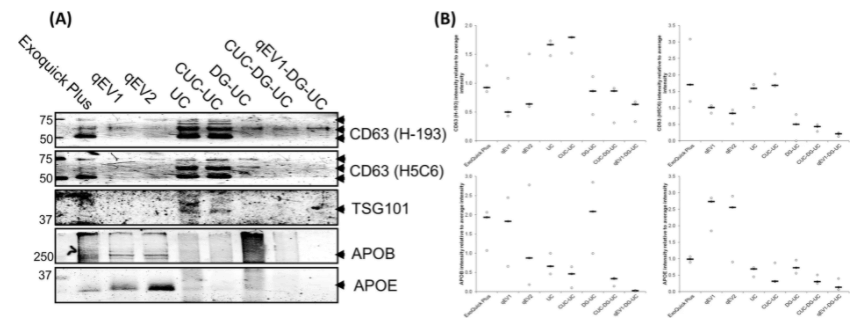 Fig 4. Western blot analysis of EV markers, CD63 and TSG101, and lipoprotein markers, APOB, and APOE. (Brennan K, et al. 2020)
Creative Bioarray has established a mature service process with years of experience in providing exosomes research products and services. You can rely on our expertise to meet all your exosomes characterization needs. You will benefit from our technical expertise and state-of-the-art facilities. If you are interested in our services or have any specific needs, please contact us. We look forward to working with you in the near future.
References:
- Brennan K, Martin K, FitzGerald S P, et al. A comparison of methods for the isolation and separation of extracellular vesicles from protein and lipid particles in human serum[J]. Scientific reports, 2020, 10(1): 1-13.
- Zhou M, Weber S R, Zhao Y, et al. Methods for exosome isolation and characterization[J]. Exosomes, 2020: 23-38.
For research use only. Not for any other purpose.