With state-of-the-art platform and experienced scientific team, Creative Bioarray can provide a wide selection of autophagy assays to accelerate our customer's research and drug development project.
Introduction of Autophagy
Autophagy is a dynamic intracellular degration process. It allows the orderly autophagosomic-lysosomal degradation and recycling of unnecessary or dysfuncitonal cellular components, such as abnormal protein aggregates, bulk cytoplasmic contents, and excess or damaged organelles. Microautophagy, macroautophagy and chaperone-mediated autophagy (CMA) are three major forms. They can be activated by various exogenous and endogenous stimuli.
Autophagy is a physiologically essential and highly regulated catabolic pathway. Like apoptosis and cell senescence, autophagy is critical in various cellular functions and biological processes, such as development and growth. It ensures the recycling of nutrients, defends against metabolic stress, helps maintain the cellular and organismal homeostasis, and plays an adaptive role in protecting organisms from various diseases.
Abnormal autophagy is implicated in many pathological conditions. The accumulation of cell damage and aging is a primary driver of faulty autophagy. It is found that faulty autophage is involved in the pathogenesis of many diseases, such as cancer, cardiac diseases, neuronal degeneration disorders, and infectious diseases. With a deeper understanding of the physiological functions, autophagy modulation has been considered as a potential therapeutic target. Further study of autophagy can help to reveal the pathogenesis of related diseases and develop new therapies.
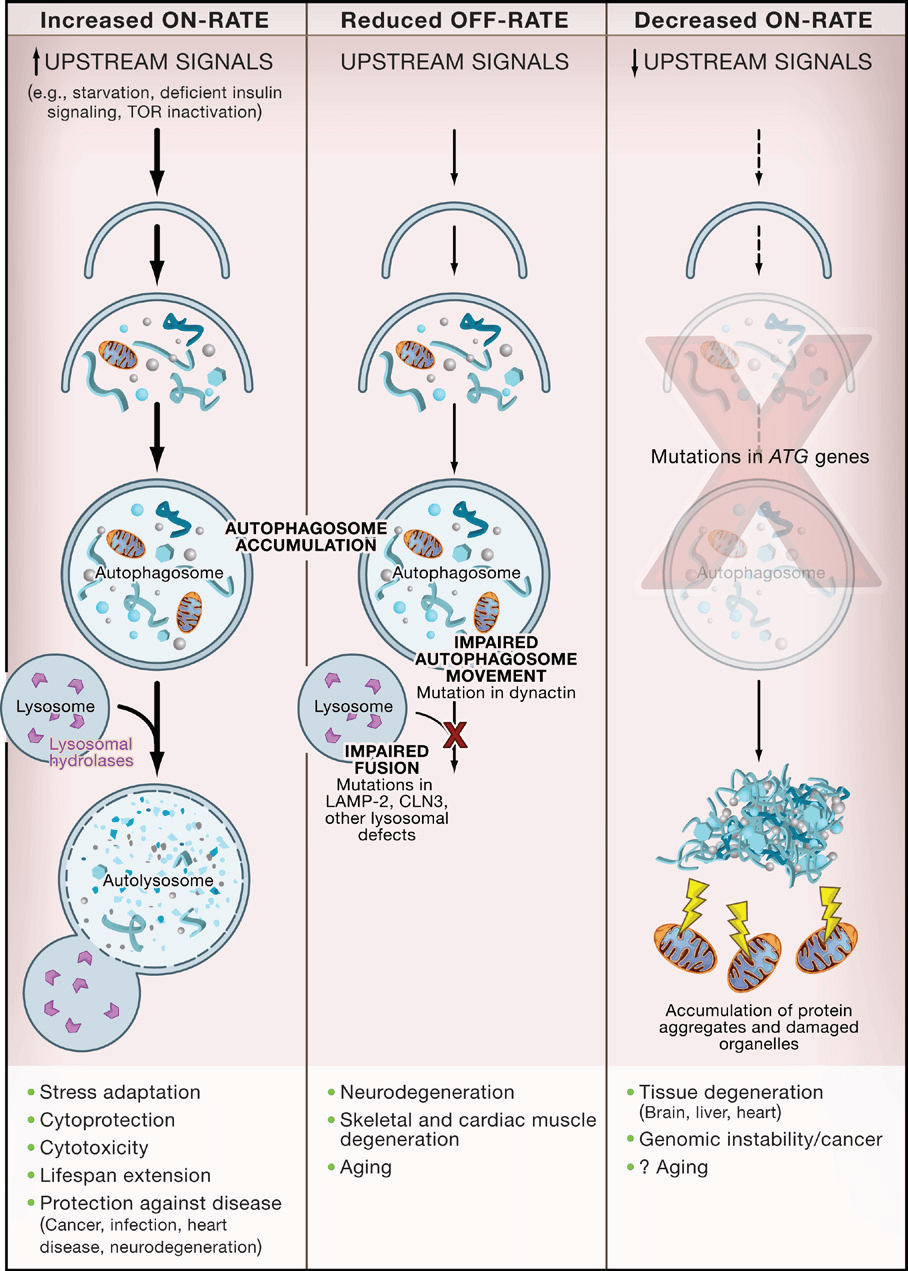
 Figure 1. Alterations in Different Stages of Autophagy Have Different Consequences. (Levine B, & Kroemer G., 2008)
Cell Autophagy Assays Available at Creative Bioarray
Direct structure observation and measurement of autophagic activity are two common approaches to monitor autophagy. We offer a wide selection of autophagy assays, including but not limited to the following:
Identification of Autophagic Structures
Autophagosomes are clear hallmarks of autophagy. Autophagosomes with double-membraned structures containing undigested cytoplamic materials can be identified by electron microscopy.
Autophagy LC3-Based Assays
Microtubule-associated protein 1A/1B-light chain 3 (LC3) is the most widely used autophagosome marker. The amount of LC3-II is associated to the number of autophagy-related structures. Detection of LC3 by immunofluorescence, immunoblotting and immunoprecipitation has become a reliable method to monitor autophagy.
GFP-LC3 is an effective tool for monitoring vacuolar/lysosomal localization and autophagic flux. p62, directly bind to LC3 can be selectively degraded by autophagy. It is another widely used marker to monitor autophagic activity.
Long-Lived Protein Degradation Assay
Measuring degradation of long-lived protein can be used to quantitatively assess bulk autophagic flux. This method is usually used in adherent cell lines and allows for high-throughput screening.
Monodansylcadaverine Staining
Monodansylcadaverine (MDC), an acidophilic autofluorescent compound, is a specific marker for autophagic vacuoles. It also can be used in vivo.
Creative Bioarray can customize the best solution according to your specific needs with fast turnaround time and competitive price. Our services are high quality and reliable. If you need more detailed information, please feel free to contact us. We look forward to cooperating with you.
References:
- Levine B, & Kroemer G. Autophagy in the pathogenesis of disease. Cell, 2008, 132(1), pp: 27-42.
- Shintani T, & Klionsky D J. Autophagy in health and disease: a double-edged sword. Science, 2004, 306(5698), pp: 990-995.
For research use only. Not for any other purpose.

 Figure 1. Alterations in Different Stages of Autophagy Have Different Consequences. (Levine B, & Kroemer G., 2008)
